
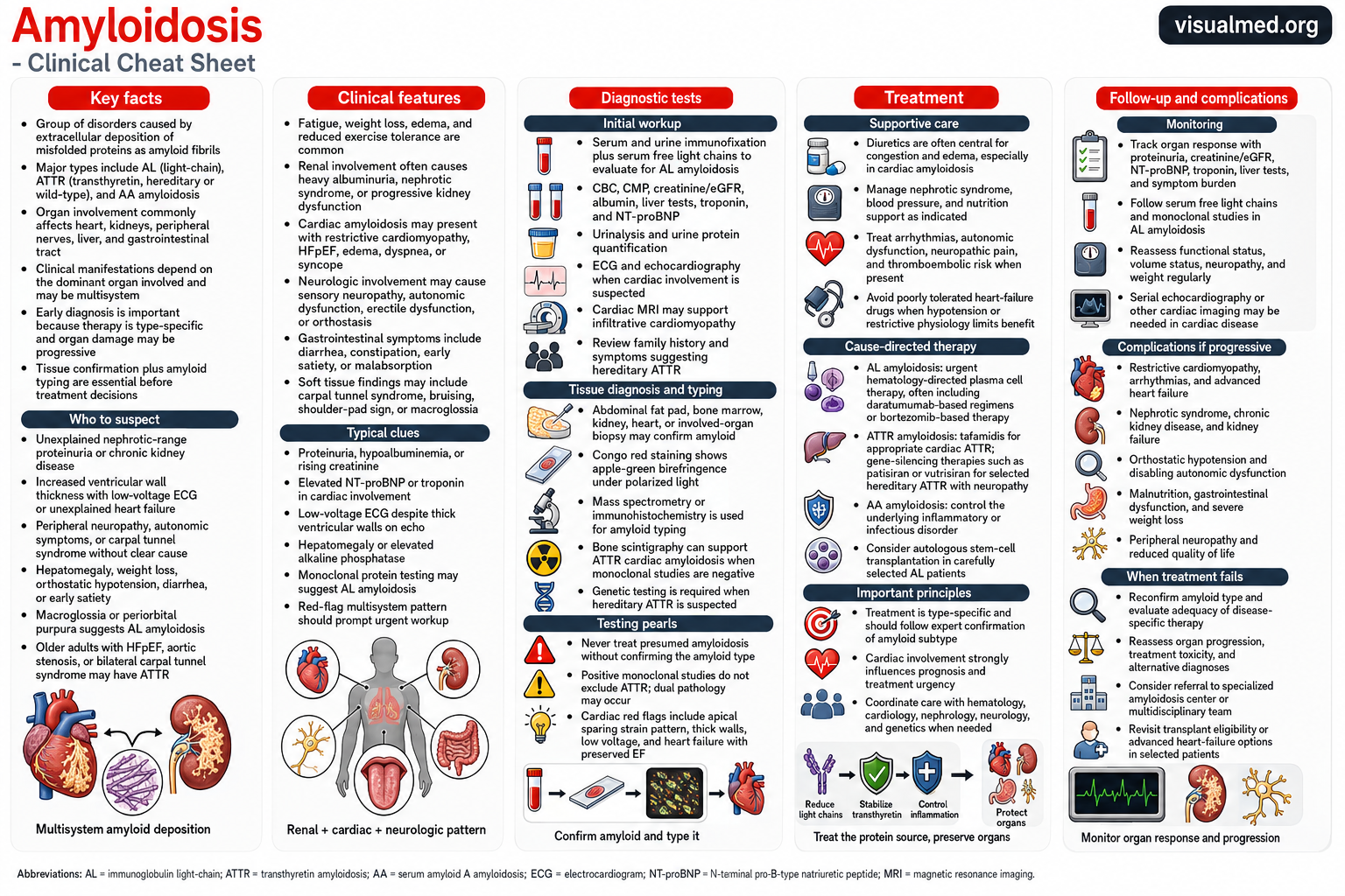
Amyloidosis comprises a heterogeneous group of disorders characterized by extracellular deposition of misfolded proteins as amyloid fibrils. The clinical phenotype depends on the precursor protein, the organs involved, and the extent of tissue deposition.
The most important systemic forms encountered in clinical practice are immunoglobulin light-chain amyloidosis, or AL amyloidosis; transthyretin amyloidosis, or ATTR amyloidosis; and serum amyloid A–associated AA amyloidosis. Cardiac, renal, neurologic, gastrointestinal, hepatic, and soft-tissue manifestations frequently overlap, making early recognition dependent on identifying a multisystem pattern rather than an isolated abnormality.
The central diagnostic principle is simple:
Amyloidosis should not be treated until the amyloid precursor protein has been identified.
Therapy is type-specific, and an incorrect assumption about amyloid subtype can lead to ineffective or potentially harmful treatment.
Major Types of Systemic Amyloidosis
AL amyloidosis
AL amyloidosis results from a clonal plasma-cell disorder that produces misfolded immunoglobulin light chains. These light chains can deposit in the heart, kidneys, peripheral nerves, liver, gastrointestinal tract, and soft tissues.
The plasma-cell clone may be relatively small, so the absence of overt multiple myeloma does not exclude AL amyloidosis. Cardiac involvement is particularly important because it strongly influences treatment tolerance, prognosis, and urgency.
ATTR amyloidosis
ATTR amyloidosis is caused by misfolding of transthyretin, a protein primarily synthesized by the liver.
It occurs in two major forms:
- Wild-type ATTR, caused by deposition of genetically normal transthyretin and predominantly associated with cardiomyopathy in older adults
- Variant or hereditary ATTR, caused by a pathogenic TTR gene variant and associated with variable combinations of cardiomyopathy, polyneuropathy, autonomic dysfunction, and other manifestations
Genetic testing is required after an ATTR diagnosis to distinguish wild-type from hereditary disease, even when the patient does not report a family history.
AA amyloidosis
AA amyloidosis develops in the setting of persistent elevation of serum amyloid A due to chronic inflammatory or infectious disease. The kidneys are commonly affected, with proteinuria, nephrotic syndrome, and progressive kidney dysfunction being frequent presentations.
Management centers on suppressing the underlying inflammatory process and reducing ongoing production of serum amyloid A.
Other systemic and localized amyloid disorders exist, including leukocyte chemotaxin-2 amyloidosis, dialysis-related beta-2 microglobulin amyloidosis, and organ-limited forms. These require separate diagnostic and therapeutic considerations.
When Should Clinicians Suspect Amyloidosis?
Amyloidosis is often missed because each individual manifestation has a broad differential diagnosis. Suspicion should increase when findings from several organ systems occur together.
Cardiac red flags
Consider cardiac amyloidosis in patients with:
- Increased ventricular wall thickness without sufficient hypertension or valvular disease to explain it
- Heart failure with preserved or mildly reduced ejection fraction
- Restrictive physiology or severe diastolic dysfunction
- Biatrial enlargement
- Atrial fibrillation or conduction-system disease
- Elevated troponin or natriuretic peptides out of proportion to the apparent clinical state
- Reduced global longitudinal strain with relative apical sparing
- Low QRS voltage relative to the degree of ventricular thickening
- Recurrent intolerance of conventional heart-failure medications
- Unexplained pericardial effusion
Low ECG voltage is not required and occurs in only a minority of patients. Its absence should not lower suspicion when the clinical and imaging phenotype is otherwise compelling.
Extracardiac clues
Important extracardiac clues include:
- Nephrotic-range proteinuria or otherwise unexplained albuminuria
- Peripheral or autonomic neuropathy
- Orthostatic hypotension
- Bilateral carpal tunnel syndrome
- Lumbar spinal stenosis
- Spontaneous distal biceps-tendon rupture
- Gastrointestinal dysmotility, diarrhea, constipation, or early satiety
- Unexplained hepatomegaly or elevated alkaline phosphatase
- Easy bruising
- Macroglossia
- Periorbital purpura
Macroglossia and periorbital purpura are particularly suggestive of AL amyloidosis, while spinal stenosis and spontaneous biceps rupture are more characteristic of ATTR.
Organ-Specific Clinical Manifestations
Renal amyloidosis
Renal involvement commonly presents with albuminuria, nephrotic-range proteinuria, hypoalbuminemia, edema, and progressive chronic kidney disease. The urine sediment may be relatively bland.
AL and AA amyloidosis are major causes of renal amyloid disease, although other precursor proteins may also involve the kidney. Proteinuria should be quantified, and the evaluation should include serum albumin, creatinine, eGFR, electrolytes, lipid profile, and assessment for nephrotic complications.
Cardiac amyloidosis
Amyloid infiltration causes increased myocardial stiffness, impaired ventricular filling, reduced stroke volume, atrial dysfunction, and progressive heart failure. Patients may develop syncope, atrial arrhythmias, conduction disease, right-sided congestion, and low-output symptoms despite a preserved ejection fraction.
Cardiac involvement in AL amyloidosis may progress rapidly and requires urgent hematologic evaluation. ATTR cardiomyopathy generally follows a more indolent course but may be substantially advanced by the time it is recognized.
Neurologic involvement
Hereditary ATTR commonly causes a length-dependent sensorimotor polyneuropathy, although AL amyloidosis can produce a similar phenotype. Autonomic manifestations may include orthostatic hypotension, erectile dysfunction, altered sweating, early satiety, diarrhea, constipation, and bladder dysfunction.
Carpal tunnel syndrome may precede overt cardiac or neurologic disease by several years and should be viewed as a potential early clue when bilateral or accompanied by other red flags.
Initial Diagnostic Evaluation
The diagnostic approach should answer three questions:
- Is amyloidosis likely?
- Is a monoclonal light-chain process present?
- What is the amyloid precursor protein?
A practical initial evaluation includes:
- CBC and comprehensive metabolic panel
- Serum creatinine and eGFR
- Serum albumin and liver tests
- Urinalysis and urine protein quantification
- Troponin and NT-proBNP when cardiac involvement is possible
- ECG
- Echocardiography
- Serum free light-chain assay
- Serum immunofixation
- Urine immunofixation
Serum or urine protein electrophoresis alone is not sufficiently sensitive to exclude a monoclonal light-chain process. The recommended monoclonal screen consists of serum free light chains together with serum and urine immunofixation.
Evaluating for AL Amyloidosis
AL amyloidosis must be excluded early because it is a hematologic emergency when significant cardiac involvement is present.
A complete monoclonal evaluation includes:
- Serum free kappa and lambda light chains with ratio
- Serum immunofixation electrophoresis
- Urine immunofixation electrophoresis
Chronic kidney disease may alter absolute free light-chain values and widen the kappa-to-lambda ratio. Results must therefore be interpreted alongside immunofixation and the renal clinical context.
When a monoclonal abnormality is identified, tissue confirmation and definitive amyloid typing are generally required. A monoclonal gammopathy is common in older adults and may coexist with ATTR amyloidosis; therefore, the presence of an abnormal light-chain study does not prove that the cardiac or renal deposits are AL.
Tissue Diagnosis
Potential biopsy sites include:
- Abdominal fat pad
- Bone marrow
- Kidney
- Heart
- Liver
- Gastrointestinal tissue
- Peripheral nerve
- Another clinically involved organ
Congo red staining demonstrates amyloid deposits, which exhibit characteristic apple-green birefringence under polarized light.
A negative abdominal fat-pad biopsy does not exclude systemic amyloidosis. When suspicion remains high, biopsy of the clinically involved organ should be considered.
Amyloid Typing
Identifying amyloid on Congo red staining is not sufficient. The precursor protein must be determined.
Mass spectrometry–based proteomic analysis is generally the preferred typing method when available because it can distinguish AL, ATTR, AA, and less common forms more reliably than morphology alone.
Immunohistochemistry or immunofluorescence may assist with typing, but interpretation can be limited by nonspecific staining or incomplete antibody panels. Treatment should not be initiated on the basis of a presumed subtype when typing remains uncertain.
Nonbiopsy Diagnosis of ATTR Cardiac Amyloidosis
ATTR cardiomyopathy may be diagnosed without an endomyocardial biopsy in an appropriate clinical setting when:
- Cardiac imaging suggests amyloidosis.
- Serum free light chains and serum and urine immunofixation show no evidence of a monoclonal process.
- Technetium-labeled bone scintigraphy demonstrates significant myocardial tracer uptake, confirmed with SPECT imaging.
If the monoclonal screen is positive, radionuclide uptake cannot reliably distinguish ATTR from AL, and tissue biopsy with amyloid typing is required.
Once ATTR is established, TTR gene sequencing should be performed to distinguish wild-type from hereditary disease and to guide family counseling and cascade testing.
Cardiac Imaging
Echocardiography
Common echocardiographic findings include:
- Increased ventricular wall thickness
- Biatrial enlargement
- Restrictive filling
- Thickening of the valves and interatrial septum
- Right-ventricular free-wall thickening
- Pericardial effusion
- Reduced longitudinal strain with relative apical sparing
These findings support but do not independently establish amyloid type.
Cardiac MRI
Cardiac MRI may demonstrate diffuse or subendocardial late gadolinium enhancement, abnormal myocardial nulling, and increased extracellular volume. It is valuable for identifying an infiltrative cardiomyopathy but cannot reliably distinguish AL from ATTR without additional testing.
Treatment Principles
Amyloidosis treatment has two simultaneous objectives:
- Reduce or eliminate production of the amyloid-forming precursor protein
- Manage existing organ dysfunction and prevent additional complications
Treatment should be coordinated through a multidisciplinary team that may include hematology, cardiology, nephrology, neurology, gastroenterology, genetics, pathology, and transplant specialists.
Supportive Management
Volume management
Loop diuretics are central to treating cardiac congestion and nephrotic edema. Combination diuretic therapy may be necessary, but overdiuresis can produce marked hypotension or renal dysfunction because patients with restrictive physiology often have a narrow preload window.
Daily weights, renal function, serum electrolytes, blood pressure, and orthostatic symptoms should guide adjustment.
Conventional heart-failure medications
Beta-blockers, ACE inhibitors, angiotensin-receptor blockers, and related vasodilating therapies may be poorly tolerated in advanced cardiac amyloidosis because patients often depend on heart rate and filling pressure to maintain cardiac output.
These medications are not universally contraindicated, but they should be introduced cautiously and continued only when clearly tolerated and clinically indicated.
Atrial fibrillation and thromboembolism
Amyloid cardiomyopathy is associated with atrial mechanical dysfunction and a substantial risk of intracardiac thrombus. The ACC consensus recommends anticoagulation for atrial fibrillation regardless of the CHA₂DS₂-VASc score, provided bleeding risk is acceptable.
Transesophageal echocardiography should be considered before cardioversion even in apparently adequately anticoagulated patients because atrial thrombi may still occur.
Autonomic dysfunction
Orthostatic hypotension may require medication review, compression garments, cautious volume management, and selected pharmacologic therapy. Management is often difficult when autonomic hypotension coexists with cardiac or renal congestion.
Treatment of AL Amyloidosis
AL treatment aims to suppress the abnormal plasma-cell clone rapidly and achieve a deep hematologic response before irreversible organ failure develops.
For newly diagnosed AL amyloidosis, daratumumab and hyaluronidase-fihj combined with bortezomib, cyclophosphamide, and dexamethasone is an FDA-approved regimen. In November 2025, the FDA converted the indication to traditional approval after longer-term ANDROMEDA data demonstrated improvements in major organ deterioration–progression-free survival and overall survival.
Treatment intensity must be adjusted to cardiac stage, frailty, blood pressure, renal function, neuropathy, and treatment tolerance. The daratumumab regimen is not recommended outside clinical trials for patients with NYHA class IIIB or IV cardiac disease or Mayo stage IIIB AL amyloidosis according to the prescribing information.
Autologous stem-cell transplantation remains an option for carefully selected patients with adequate functional status and limited advanced cardiac involvement. Selection should occur at an experienced amyloidosis center because transplant-related risk differs substantially from that in conventional multiple myeloma.
The hematologic response is followed using the involved and uninvolved free light chains, immunofixation, and bone-marrow or clonal assessments when indicated. Organ improvement often lags behind hematologic response.
Treatment of ATTR Amyloidosis
ATTR therapies either stabilize circulating transthyretin or reduce its hepatic production.
TTR stabilizers
Tafamidis is an oral TTR stabilizer approved for wild-type or hereditary ATTR cardiomyopathy to reduce cardiovascular mortality and cardiovascular-related hospitalization.
Acoramidis is another oral TTR stabilizer approved in 2024 for adults with wild-type or variant ATTR cardiomyopathy to reduce cardiovascular death and cardiovascular-related hospitalization.
TTR silencers
Vutrisiran is a small-interfering RNA therapy that suppresses hepatic TTR synthesis. It is FDA-approved for hereditary ATTR polyneuropathy and, since 2025, for wild-type or hereditary ATTR cardiomyopathy to reduce cardiovascular mortality, cardiovascular hospitalizations, and urgent heart-failure visits.
Other gene-silencing therapies may be used for hereditary ATTR polyneuropathy depending on indication, availability, phenotype, comorbidities, and regulatory jurisdiction.
Disease-modifying therapy is most effective before advanced irreversible organ dysfunction develops. Treatment does not remove all pre-existing deposits immediately, and clinical improvement may be gradual.
Treatment of AA Amyloidosis
The therapeutic priority in AA amyloidosis is sustained suppression of the inflammatory or infectious driver.
Depending on the cause, this may involve:
- Biologic or conventional anti-inflammatory therapy
- Treatment of chronic infection
- Colchicine for familial Mediterranean fever
- Control of inflammatory arthritis or autoinflammatory disease
- Surgical or procedural source control when appropriate
Renal supportive care includes control of blood pressure and proteinuria, management of edema, and treatment of progressive kidney failure.
Monitoring Treatment Response
Monitoring must be tailored to amyloid type and the organs involved.
AL amyloidosis
Follow:
- Serum free light chains and difference between involved and uninvolved chains
- Serum and urine immunofixation
- NT-proBNP and troponin
- Proteinuria and serum albumin
- Creatinine and eGFR
- Liver tests
- Functional status and symptom burden
A hematologic response may occur before cardiac or renal improvement. Rising free light chains may precede organ progression and should prompt reassessment of clonal control.
ATTR amyloidosis
Follow:
- Heart-failure symptoms and functional capacity
- Volume status and diuretic requirements
- NT-proBNP and troponin trends
- ECG and rhythm monitoring when appropriate
- Echocardiography
- Neurologic function in hereditary disease
- Nutrition and body weight
- Treatment-specific safety parameters
For patients receiving vutrisiran, vitamin A supplementation at the recommended daily allowance is advised because the medication reduces circulating vitamin A levels.
Renal disease
Follow:
- Urine protein excretion
- Serum albumin
- Creatinine and eGFR
- Edema and body weight
- Blood pressure
- Thrombotic and infectious complications of nephrotic syndrome
Major Complications
Progressive systemic amyloidosis may lead to:
- Restrictive cardiomyopathy and advanced heart failure
- Atrial and ventricular arrhythmias
- Conduction disease
- Intracardiac thrombus and systemic embolism
- Nephrotic syndrome
- Progressive chronic kidney disease and kidney failure
- Severe orthostatic hypotension
- Peripheral and autonomic neuropathy
- Gastrointestinal dysmotility and malnutrition
- Hepatic dysfunction
- Treatment-related infection or cytopenias
Cardiac involvement is among the strongest determinants of prognosis, particularly in AL amyloidosis.
Common Diagnostic Pitfalls
Assuming thick ventricular walls equal hypertensive heart disease
Amyloidosis should remain in the differential when ventricular thickening is accompanied by disproportionate diastolic dysfunction, reduced longitudinal strain, conduction disease, low blood pressure, neuropathy, proteinuria, or carpal tunnel syndrome.
Using SPEP alone to exclude AL
Serum electrophoresis alone is inadequate. Serum free light chains plus serum and urine immunofixation are required for an appropriate monoclonal screen.
Diagnosing ATTR from a positive scan despite a monoclonal gammopathy
Bone scintigraphy can support a nonbiopsy ATTR diagnosis only after an adequate monoclonal evaluation is negative. A positive scan in a patient with a monoclonal protein requires tissue confirmation and typing.
Treating an untyped Congo red–positive biopsy
Congo red confirms amyloid but does not identify the precursor protein. Treatment should wait for reliable typing.
Considering a negative fat-pad biopsy definitive
A negative surrogate biopsy does not exclude systemic disease. Biopsy of an involved organ may be necessary when clinical suspicion remains high.
Key Takeaway
Amyloidosis should be suspected when otherwise unexplained cardiac thickening, HFpEF, nephrotic proteinuria, neuropathy, autonomic dysfunction, carpal tunnel syndrome, or characteristic soft-tissue findings occur—especially in combination.
The clinician’s essential workflow is:
- Recognize the multisystem pattern.
- Perform serum free light chains plus serum and urine immunofixation.
- Confirm amyloid when necessary with tissue biopsy.
- Determine the precursor protein using definitive typing.
- Use bone scintigraphy for nonbiopsy ATTR diagnosis only when AL has been excluded.
- Initiate type-specific treatment promptly.
- Monitor both precursor-protein response and organ response.
Early and accurate typing now matters more than ever because effective disease-modifying therapies are available for both AL and ATTR amyloidosis.
This article is intended for clinician education and should be applied alongside specialist consultation, current prescribing information, institutional protocols, and individualized clinical judgment.