
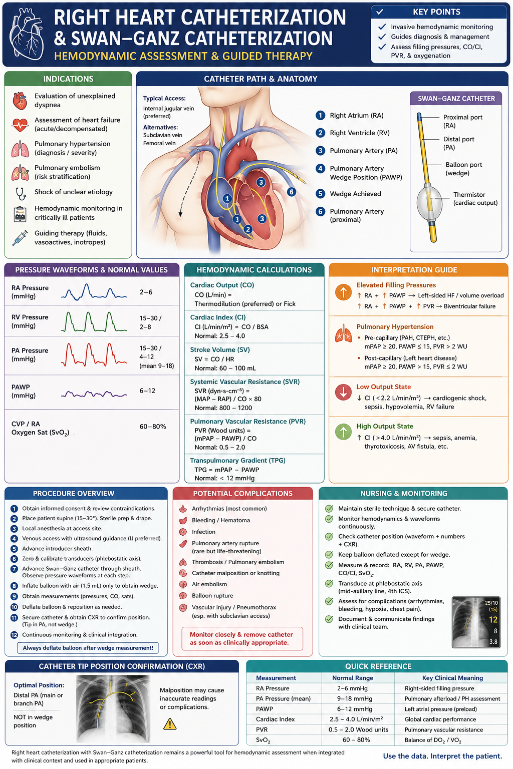
A clinician-focused review of right heart catheterization and Swan-Ganz catheterization, including indications, catheter positioning, pressure waveforms, cardiac-output measurement, pulmonary hypertension classification, shock profiles, complications, and common interpretation errors.
Right heart catheterization, or RHC, provides direct measurement of right-sided and pulmonary hemodynamics. It remains the reference standard for confirming pulmonary hypertension, differentiating pre- from post-capillary physiology, evaluating selected patients with shock or advanced heart failure, and resolving clinical situations in which noninvasive findings are inconclusive.
A Swan-Ganz catheter, also called a pulmonary artery catheter, is a balloon-tipped, flow-directed catheter used during RHC. The terms are related but not interchangeable: RHC describes the invasive procedure, while the Swan-Ganz catheter is one device that can be used to obtain measurements and, when clinically justified, remain in place for serial monitoring. Expert consensus emphasizes defining the clinical question before catheterization and integrating the numbers with the patient’s examination, imaging, and treatment response.
When Is Right Heart Catheterization Indicated?
Common clinical indications include:
- Confirmation and classification of suspected pulmonary hypertension
- Unexplained dyspnea when noninvasive testing is inconclusive
- Cardiogenic shock or mixed shock with uncertain hemodynamics
- Advanced or decompensated heart failure with unclear filling pressures
- Assessment before heart or lung transplantation
- Evaluation of intracardiac shunts
- Differentiation of constrictive pericarditis from restrictive cardiomyopathy
- Assessment of selected valvular or congenital heart diseases
- Guidance of fluids, vasopressors, inotropes, diuretics, or mechanical circulatory support
Pulmonary hypertension cannot be definitively classified from echocardiography alone. Current ESC/ERS guidance bases the hemodynamic definition of pulmonary hypertension on measurements obtained during RHC, interpreted within the broader clinical context.
In cardiogenic shock, pulmonary artery catheterization may help establish the shock phenotype, quantify left- and right-sided filling pressures, assess cardiac output, and guide escalation or de-escalation of vasoactive and mechanical support. The 2025 ACC cardiogenic-shock guidance specifically includes invasive pulmonary artery catheter monitoring as a tool for diagnosis and management.
RHC should not be used reflexively in every patient with routine decompensated heart failure. Its value is greatest when clinical assessment is inadequate, the hemodynamic profile is uncertain, symptoms persist despite empiric therapy, or management depends on accurately distinguishing congestion, low output, pulmonary vascular disease, or right-ventricular failure.
Vascular Access and Catheter Path
Venous access is generally obtained through the:
- Internal jugular vein
- Femoral vein
- Subclavian vein
- Selected upper-extremity veins in appropriate cases
The right internal jugular vein frequently provides a relatively direct route to the right heart. Ultrasound-guided vascular access is preferred when feasible because it improves access accuracy and can reduce mechanical complications. Contemporary cath-lab best-practice guidance emphasizes both vascular ultrasound and procedure-specific access planning.
The balloon-tipped catheter is advanced sequentially through:
- Right atrium
- Right ventricle
- Pulmonary artery
- A distal pulmonary arterial branch to obtain the pulmonary artery wedge pressure
The operator should recognize the corresponding waveform at each stage. Fluoroscopy may be used to confirm catheter course and position, particularly when advancement is difficult or the anatomy is abnormal.
The balloon should remain deflated except during flow-directed advancement or brief wedge acquisition. Prolonged inflation or distal catheter migration increases the risk of pulmonary infarction or pulmonary artery injury.
Transducer Setup and Signal Quality
Accurate measurements begin before the catheter enters the heart.
The pressure transducer should be:
- Zeroed to atmospheric pressure
- Leveled at the approximate left-atrial position
- Checked for air bubbles, loose connections, clot, or excessive tubing
- Reassessed when the patient’s position changes
Pressure values should be interpreted only after waveform morphology and signal fidelity have been confirmed. A plausible-looking number derived from an overdamped, underdamped, unzeroed, or incorrectly positioned system may be more misleading than no measurement at all.
Respiratory variation must also be considered. In many patients, pressures are recorded at end expiration, when intrathoracic pressure most closely approximates atmospheric pressure. In patients with marked respiratory effort, obesity, obstructive lung disease, or positive-pressure ventilation, relying on a single automated value may misclassify the hemodynamic profile. Measurements should be reviewed directly from the tracing and interpreted in the respiratory and ventilatory context.
Core Pressure Measurements
Right-atrial pressure
Right-atrial pressure reflects right-sided filling pressure and contains characteristic a and v waves.
An elevated value may occur with:
- Right-ventricular failure
- Tricuspid regurgitation
- Volume overload
- Constrictive pericarditis
- Restrictive cardiomyopathy
- Pulmonary hypertension
Large v waves may suggest significant tricuspid regurgitation, although waveform interpretation must account for catheter position, rhythm, and pressure transmission.
Right-ventricular pressure
Right-ventricular systolic pressure normally resembles pulmonary artery systolic pressure in the absence of pulmonic-valve or right-ventricular outflow obstruction. Right-ventricular end-diastolic pressure generally tracks right-atrial pressure.
A marked systolic gradient between the right ventricle and pulmonary artery suggests pulmonic stenosis or dynamic or fixed right-ventricular outflow obstruction.
The catheter should not remain unnecessarily in the right ventricle because mechanical irritation commonly produces premature ventricular contractions or nonsustained ventricular tachycardia.
Pulmonary artery pressure
The pulmonary artery tracing has a systolic peak, diastolic pressure, and dicrotic notch caused by pulmonic-valve closure.
The mean pulmonary artery pressure should be derived by electronic integration of the waveform rather than estimated simply as the arithmetic average of systolic and diastolic pressures.
Pulmonary artery wedge pressure
Pulmonary artery wedge pressure, or PAWP, estimates left-atrial pressure when a pulmonary arterial branch is fully occluded and the catheter is connected through an uninterrupted static blood column to the pulmonary veins and left atrium.
A valid wedge tracing should demonstrate:
- Atrial waveform morphology
- Lower pulsatility than the pulmonary artery tracing
- An appropriate pressure relationship to pulmonary artery diastolic pressure
- A position that is neither excessively proximal nor dangerously distal
When the tracing is uncertain, confirmation may include fluoroscopic review and sampling blood from the distal port. A truly wedged sample should have oxygen saturation approximating pulmonary venous or systemic arterial blood.
PAWP can be misleading in severe mitral regurgitation, mitral stenosis, pulmonary venous obstruction, high intrathoracic pressure, partial wedging, or lung-zone conditions that interrupt the expected relationship between alveolar and vascular pressure.
Cardiac Output and Cardiac Index
Cardiac output is commonly measured by:
- Thermodilution
- Direct Fick calculation
Thermodilution uses the temperature change produced by injection of a known volume of fluid through the proximal catheter port. Several technically acceptable measurements are averaged. Results may be less reliable with severe tricuspid regurgitation, intracardiac shunting, very low output, or inconsistent injection technique.
The direct Fick method calculates cardiac output from measured oxygen consumption and the arterial-to-mixed venous oxygen-content difference:
Cardiac output = oxygen consumption ÷ arteriovenous oxygen-content difference
Directly measured oxygen consumption is preferable when precision is essential. “Estimated Fick” methods use assumed oxygen consumption and may introduce substantial error in patients with obesity, critical illness, abnormal metabolic states, or unusual body composition.
Cardiac index adjusts cardiac output for body surface area:
Cardiac index = cardiac output ÷ body surface area
A low cardiac index is not a diagnosis by itself. It must be interpreted with blood pressure, filling pressures, systemic vascular resistance, lactate, urine output, mental status, and other markers of tissue perfusion.
Derived Hemodynamic Calculations
Pulmonary vascular resistance
PVR = (mean pulmonary artery pressure − PAWP) ÷ cardiac output
The result is expressed in Wood units.
PVR represents the pressure drop across the pulmonary circulation relative to flow. It should not be interpreted without confirming that the mean pulmonary pressure, wedge pressure, and cardiac output were all accurately obtained under the same physiologic conditions.
Systemic vascular resistance
SVR = (mean arterial pressure − right-atrial pressure) ÷ cardiac output × 80
The result is expressed in dynes·seconds·cm⁻⁵.
A low SVR may support distributive physiology, whereas a high SVR may reflect vasoconstriction, low-output compensation, or vasoactive therapy.
Transpulmonary gradient
TPG = mean pulmonary artery pressure − PAWP
TPG reflects the total pressure difference across the pulmonary circulation but is influenced by flow and left-atrial pressure.
Diastolic pulmonary gradient
DPG = pulmonary artery diastolic pressure − PAWP
DPG may provide additional context but should not be used in isolation to classify disease or make treatment decisions.
Hemodynamic Definition of Pulmonary Hypertension
The contemporary hemodynamic definition of pulmonary hypertension is:
Mean pulmonary artery pressure greater than 20 mm Hg
The main invasive categories are:
Pre-capillary pulmonary hypertension
- Mean pulmonary artery pressure >20 mm Hg
- PAWP ≤15 mm Hg
- PVR >2 Wood units
This pattern may occur with pulmonary arterial hypertension, chronic thromboembolic pulmonary hypertension, or pulmonary hypertension associated with lung disease.
Isolated post-capillary pulmonary hypertension
- Mean pulmonary artery pressure >20 mm Hg
- PAWP >15 mm Hg
- PVR ≤2 Wood units
This profile generally supports pulmonary hypertension related predominantly to left-heart disease.
Combined pre- and post-capillary pulmonary hypertension
- Mean pulmonary artery pressure >20 mm Hg
- PAWP >15 mm Hg
- PVR >2 Wood units
This pattern indicates elevated left-sided filling pressure with an additional pulmonary vascular component.
These cutoffs should not replace clinical classification. A patient’s diagnosis still depends on imaging, pulmonary testing, thromboembolic evaluation, left-heart disease, medications, and associated systemic conditions.
Hemodynamic Profiles in Shock
RHC can help distinguish several shock phenotypes:
Predominantly left-ventricular cardiogenic shock
Typical findings include:
- Reduced cardiac index
- Elevated PAWP
- Elevated systemic vascular resistance before or during vasopressor therapy
- Variable right-atrial pressure
Right-ventricular failure
Common findings include:
- Elevated right-atrial pressure
- Disproportionately lower PAWP
- Reduced cardiac index
- Reduced pulmonary artery pulsatility
- Elevated right-atrial pressure-to-PAWP relationship
Hypovolemic physiology
Typical findings include:
- Low right-atrial pressure
- Low PAWP
- Reduced cardiac output
- Compensatory elevation in SVR
Distributive physiology
Typical findings include:
- Low SVR
- Normal or increased cardiac output early in the course
- Variable filling pressures
Mixed profiles are common, particularly in critically ill patients receiving positive-pressure ventilation, vasopressors, inotropes, diuretics, or mechanical support. The catheter should be used to follow physiology, not merely to assign a one-time label.
Intracardiac Shunt Assessment
When a shunt is suspected, oxygen saturations can be sampled sequentially from the venae cavae, right atrium, right ventricle, pulmonary artery, and systemic arterial circulation.
A reproducible oxygen-saturation step-up may localize a left-to-right shunt:
- Right-atrial step-up: atrial-level shunt
- Right-ventricular step-up: ventricular-level shunt
- Pulmonary-artery step-up: great-vessel or ductal-level shunt
Pulmonary-to-systemic flow ratio, or Qp:Qs, can then be calculated using oxygen-content relationships. Samples should be collected systematically because supplemental oxygen, sedation, fluctuating cardiac output, and inconsistent timing can create misleading results.
Provocative Hemodynamic Testing
Resting measurements may appear normal despite exertional symptoms. Selected patients may therefore undergo:
- Supine exercise hemodynamics
- Fluid challenge
- Vasodilator testing for specific pulmonary arterial hypertension indications
Exercise or fluid challenge can uncover abnormal increases in PAWP consistent with occult left-heart disease. These tests require standardized protocols and experienced interpretation.
Acute pulmonary vasoreactivity testing is not a routine test for every form of pulmonary hypertension. It is primarily used in selected patients with idiopathic, heritable, or drug-associated pulmonary arterial hypertension to identify the small subgroup that may respond to high-dose calcium-channel blocker therapy.
Potential Complications
Complications include:
- Venous bleeding or hematoma
- Pneumothorax or hemothorax
- Infection
- Venous thrombosis
- Air embolism
- Right bundle-branch block
- Complete heart block in a patient with pre-existing left bundle-branch block
- Atrial or ventricular arrhythmias
- Catheter knotting or entrapment
- Pulmonary infarction
- Balloon rupture
- Pulmonary artery perforation or rupture
Pulmonary artery rupture is uncommon but potentially catastrophic. Risk rises with advanced age, pulmonary hypertension, anticoagulation, distal catheter position, and excessive balloon inflation.
Common Interpretation Errors
Trusting the number without inspecting the tracing
Every pressure should be validated against waveform morphology, respiratory phase, transducer level, and catheter position.
Accepting an incomplete wedge
A partial wedge can falsely elevate the recorded pressure and lead to incorrect classification as post-capillary pulmonary hypertension.
Using estimated Fick output uncritically
Assumed oxygen consumption may materially distort cardiac output and PVR.
Measuring values under changing conditions
Hemodynamics obtained before and after sedation, oxygen, vasopressors, diuresis, or mechanical ventilation may not be directly comparable.
Ignoring large v waves
Mean PAWP may be substantially increased by large v waves. The tracing should be interpreted in relation to mitral regurgitation and the specific clinical question.
Treating pulmonary artery catheter targets rather than the patient
There is no universally optimal filling pressure or cardiac index for every clinical situation. The goal is adequate perfusion and controlled congestion without provoking ischemia, hypotension, renal injury, or right-heart deterioration.
Key Takeaway
Right heart catheterization is most useful when it answers a specific question that noninvasive assessment cannot resolve.
The clinician’s practical sequence is:
- Define the diagnostic or management objective.
- Obtain technically valid waveforms.
- Confirm PAWP and cardiac output rather than accepting automated values.
- Calculate derived variables from measurements obtained under stable conditions.
- Integrate the hemodynamics with imaging and the clinical phenotype.
- Reassess after treatment when serial data will change management.
- Remove an indwelling catheter as soon as its benefits no longer outweigh its risks.
This article is intended for clinician education and should be applied alongside specialist guidance, institutional protocols, and individualized clinical judgment.