
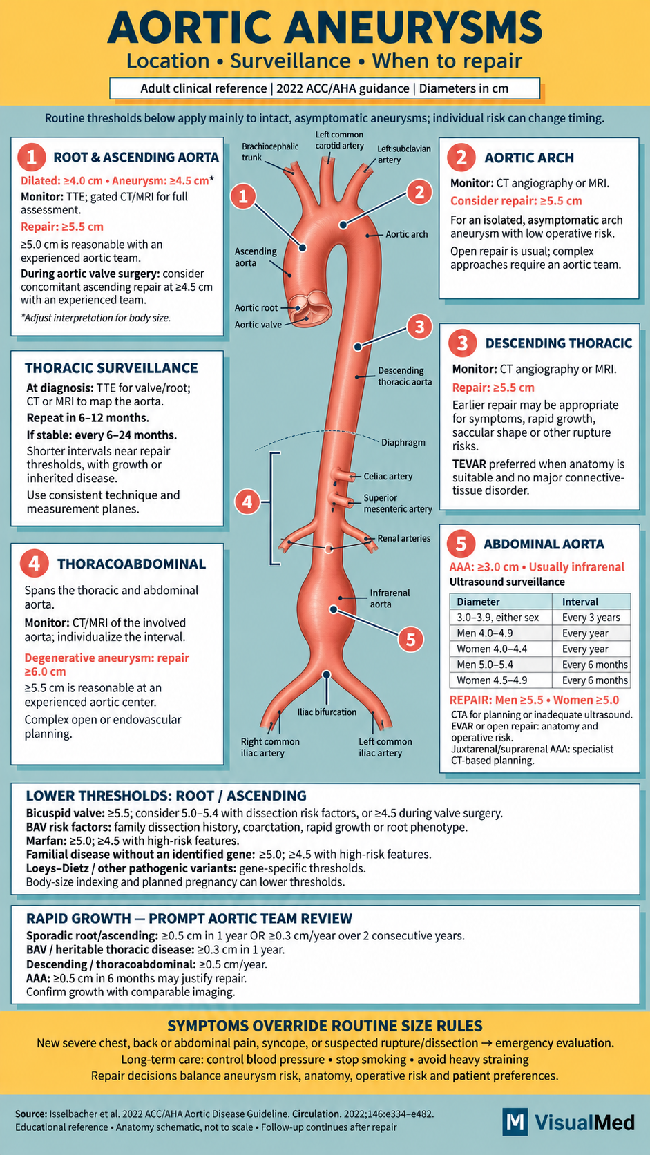
An aortic aneurysm is an abnormal enlargement of the body’s main artery. Many aneurysms develop without symptoms, making imaging essential for detecting them and monitoring their growth. The goal is to identify patients who need repair before a life-threatening rupture or dissection occurs.
Management depends on more than size alone. Location, growth rate, symptoms, valve anatomy, inherited conditions, and surgical risk all influence the decision.
This guide accompanies the VisualMed infographic and summarizes the 2022 ACC/AHA aortic disease guideline. The thresholds below primarily apply to intact, asymptomatic adult aneurysms.
Understanding the Different Aortic Locations
The aorta begins at the aortic valve, travels upward, curves through the chest, and descends into the abdomen before dividing into the common iliac arteries.
| Segment | Location |
|---|---|
| Aortic root | Surrounds the aortic valve and contains the origins of the coronary arteries. |
| Ascending aorta | Travels upward from the root toward the arch. |
| Aortic arch | Curves across the chest and supplies the major arteries to the head and arms. |
| Descending thoracic aorta | Extends from beyond the left subclavian artery to the diaphragm. |
| Abdominal aorta | Extends from the diaphragm to the iliac bifurcation. |
| Thoracoabdominal aneurysm | Involves both the thoracic and abdominal portions of the aorta. |
Most abdominal aortic aneurysms are infrarenal, meaning they occur below the arteries supplying the kidneys. Aneurysms involving or approaching the renal arteries require more complex repair planning.
What Size Is Considered an Aneurysm?
For many adults of average body size, the root or ascending aorta is considered dilated at ≥4.0 cm and aneurysmal at ≥4.5 cm. These values require adjustment for body size and should not be applied to every aortic segment.
An abdominal aortic aneurysm, or AAA, is generally defined as ≥3.0 cm. Being diagnosed with an aneurysm does not automatically mean surgery is needed. Many smaller aneurysms are managed with surveillance and treatment of cardiovascular risk factors.
How Often Should Thoracic Aneurysms Be Monitored?
At diagnosis, an echocardiogram evaluates the aortic valve, root, and visible ascending aorta. CT or MRI helps assess portions that echocardiography cannot adequately visualize.
For thoracic aortic dilation, follow-up imaging is generally reasonable:
- After 6–12 months to establish the growth rate.
- Every 6–24 months if stable, depending on diameter and individual risk.
Patients approaching a repair threshold or with concerning growth or inherited disease may need closer follow-up. Arch and descending aneurysms usually require CT or MRI.
Consistent measurement technique matters. A small difference between an echocardiogram and a CT scan should be reviewed before being labeled true growth.
Abdominal Aortic Aneurysm Surveillance
Ultrasound is usually the preferred test for monitoring uncomplicated infrarenal AAA. Surveillance intervals differ by diameter and sex:
| AAA diameter | Patient group | Usual imaging interval |
|---|---|---|
| 3.0–3.9 cm | Men and women | Every 3 years |
| 4.0–4.9 cm | Men | Every 12 months |
| 4.0–4.4 cm | Women | Every 12 months |
| 5.0–5.4 cm | Men | Every 6 months |
| 4.5–4.9 cm | Women | Every 6 months |
Once the repair threshold is reached, the next step is repair assessment rather than simply continuing routine surveillance. CT is used when ultrasound is inadequate and for preoperative planning.
When Should an Aortic Aneurysm Be Repaired?
| Location | Typical elective repair threshold |
|---|---|
| Sporadic root or ascending aorta | ≥5.5 cm; repair at ≥5.0 cm is reasonable with an experienced multidisciplinary aortic team. |
| Isolated aortic arch | Repair is reasonable at ≥5.5 cm in asymptomatic patients with low operative risk. |
| Descending thoracic aorta | Repair is recommended at ≥5.5 cm. |
| Degenerative thoracoabdominal aneurysm | Repair is recommended at ≥6.0 cm; ≥5.5 cm is reasonable at an experienced center. |
| Abdominal aortic aneurysm | ≥5.5 cm in men or ≥5.0 cm in women. |
These thresholds guide elective decisions. Symptoms, rapid growth, and other high-risk features can justify intervention sooner; substantial procedural risk may favor continued observation.
For patients already undergoing aortic valve surgery, concomitant ascending aortic replacement at ≥4.5 cm may be reasonable with an experienced team. This is a specific surgical circumstance, rather than the routine threshold for all patients.
When Do Smaller Aneurysms Need Surgery?
Certain conditions warrant consideration of earlier repair:
- Bicuspid aortic valve: repair at 5.0–5.4 cm may be reasonable with risk factors such as family dissection history, coarctation, rapid growth, or root-predominant disease.
- Marfan syndrome: root repair is recommended at ≥5.0 cm; ≥4.5 cm is reasonable with high-risk features.
- Nonsyndromic heritable thoracic disease: when no gene is identified, thresholds commonly range from 4.5–5.0 cm according to family history and other risk features.
- Loeys–Dietz syndrome and other genetic aortopathies: thresholds depend on the specific gene and clinical profile.
Body-size indexing and planned pregnancy can also alter intervention thresholds.
A detailed family history is therefore essential. Selected patients need genetic testing, and family members may need screening imaging.
How Much Growth Is Concerning?
For a sporadic root or ascending aneurysm, confirmed growth of ≥0.5 cm in one year, or ≥0.3 cm annually for two consecutive years, is an indication for surgery.
For bicuspid valve or heritable thoracic disease, ≥0.3 cm in one year is considered rapid growth. For descending thoracic or thoracoabdominal aneurysms, ≥0.5 cm annually is a high-risk feature. An AAA growing ≥0.5 cm in six months may warrant repair.
Treatment Beyond Surgery
Medical management remains important throughout surveillance. Priorities include stopping smoking, controlling blood pressure, and managing cholesterol and other cardiovascular conditions.
Exercise advice should be individualized. Moderate activity may be appropriate, while heavy lifting and intense straining may be discouraged. Patients should discuss suitable activity with their treating team rather than assume all exercise is unsafe.
Open Surgery Versus Endovascular Repair
Open repair replaces the affected aortic segment with a surgical graft. Endovascular repair places a covered stent inside the artery to exclude the aneurysm from blood flow.
Root and ascending aneurysms generally require open surgery. Suitable descending thoracic aneurysms may be treated with TEVAR, while abdominal aneurysms may be treated with EVAR or open repair. Anatomy, connective-tissue disease, procedural risk, and durability influence the choice.
Follow-up continues after repair. Imaging checks the remaining aorta and identifies complications such as an endoleak, in which blood continues entering the aneurysm sac around an endovascular graft. The surveillance schedule depends on the procedure and residual disease.
Symptoms That Require Emergency Evaluation
Sudden severe chest, back, or abdominal pain—especially with fainting, marked light-headedness, or collapse—requires emergency evaluation. Call emergency services rather than waiting for a scheduled scan.
A previously reassuring diameter does not rule out an acute complication. Surveillance is designed to guide planned care; new concerning symptoms require immediate attention.
Primary reference: Isselbacher EM, et al. 2022 ACC/AHA Guideline for the Diagnosis and Management of Aortic Disease. Circulation. 2022;146–e482.